
SETTING THE RECORD STRAIGHT
Breast CT Myth-Busting
The Evolution of Dedicated Breast CT
Dedicated breast CT—also known as cone-beam breast CT—is a next-generation 3D imaging modality that captures the breast volumetrically without compression. After decades of academic research, one developer - Koning Health - successfully advanced the concept into the first FDA-approved, clinically available system for diagnostic use, making breast CT a reality for patients and clinicians.*
Today, several other groups are working on their own prototypes, but most remain in development or early evaluation. Knowing which technologies are currently available for clinical use helps physicians and patients make informed decisions.
A new chapter in 3D breast imaging

What Is Dedicated Breast CT?
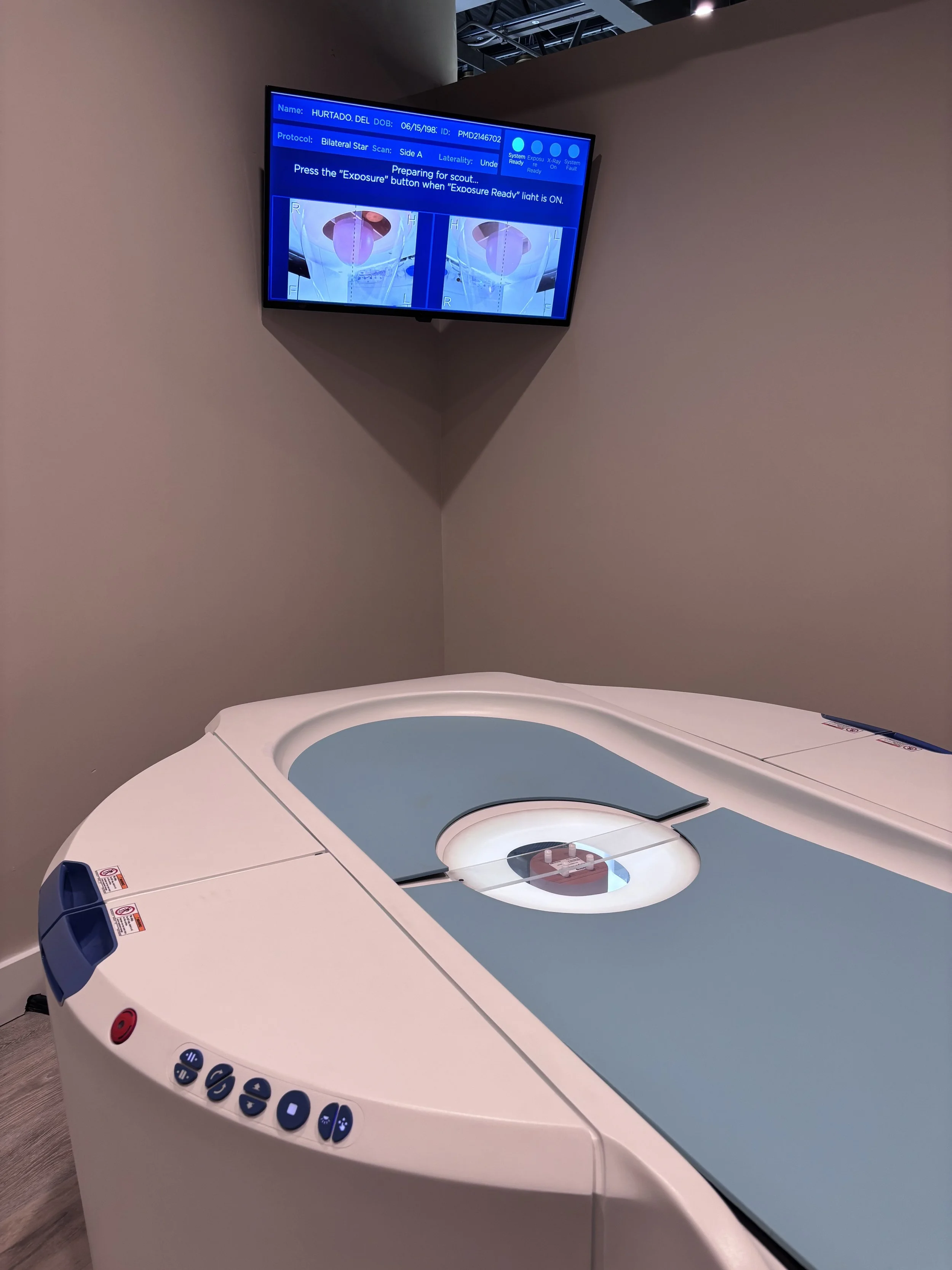
True volumetric imaging for dense tissue
Dedicated breast CT creates an isotropic, 3D dataset of the breast, allowing clinicians to visualize internal structures from every angle. The patient lies comfortably on a padded table in a prone position—no compression required—while a short rotation captures the entire breast in less than 10 seconds.
This approach provides natural separation of overlapping tissue and can be especially valuable for patients with dense breasts, where 2D methods may obscure subtle findings.

FAQs
-
FAQs -
-
Dedicated breast CT is a three-dimensional cone-beam imaging modality designed specifically for the breast.
Unlike mammography, it acquires a volumetric dataset of the entire breast in a single rotation, without the need for compression. The patient lies comfortably in a prone position while an X-ray source and detector rotate beneath the table, producing hundreds of thin slices that can be reconstructed into true 3D images. -
The scientific groundwork for breast-specific computed tomography began at the University of Rochester in the early 2000s. The inventor, Dr. Ruola Ning, was exploring how cone-beam CT geometry—already used in dental and extremity imaging—could be adapted for the breast, with careful attention to radiation dose and image quality.
Through extensive prototyping and collaboration between academic engineers and clinical radiologists, the first fully functional breast-dedicated CT scanners were tested in pilot trials. Over the following decade, this foundational research evolved into a regulatory-approved, commercially available system manufactured by Koning Health and based out of Norcross, marking the first successful transition from research concept to clinical product in this category.
This achievement effectively pioneered the dedicated breast CT field, setting performance, safety, and workflow benchmarks that later entrants have aimed to follow.
-
Imaging geometry: Breast CT performs a full 360° acquisition around the breast, rather than angled planar views.
No compression: The patient lies prone; the breast is gently suspended through the opening of the table.
True isotropic 3D data: Every voxel of the breast is captured, enabling multiplanar and volumetric visualization.
Dense tissue handling: Because there is no tissue overlap, structures obscured in 2D mammograms can appear with greater clarity when imaged volumetrically.
-
Yes. After years of research and technical validation, the first dedicated breast CT system received FDA approval for diagnostic use in the United States—becoming the inaugural commercially distributed model of its kind. That milestone established a new imaging category and demonstrated that the technology could meet clinical, engineering, and regulatory standards.
Today, a limited number of additional systems are in development or clinical evaluation, but only Koning Health manufactures the only approved model which has achieved widespread, regulated use in healthcare environments.
-
Dedicated breast CT is available in select imaging centers and hospitals in the United States and internationally. Adoption continues to grow as clinicians become familiar with the workflow, reimbursement pathways, and diagnostic benefits of true 3D breast imaging.
-
Before this technology, there was no modality capable of producing true isotropic 3D images of the entire breast at a clinically acceptable dose without compression.
The initial commercial system proved that such imaging could be achieved safely and effectively, paving the way for continued innovation in volumetric breast imaging and future imaging applications. -
Several organizations and academic groups are pursuing new versions or variations of dedicated breast CT.
All of these remain in research, prototype, or regulatory-submission stages. While their progress is promising, these systems are not yet routinely available for clinical use. The first approved and distributed breast CT system remains the reference point for clinical practice and technical comparison. This system was developed in Rochester, NY and is now manufactured in Norcross, GA. -
Dedicated breast CT uses ionizing radiation, similar in principle to other X-ray–based modalities. The dose is managed under physician oversight and designed to stay within diagnostic reference levels. Each exam is performed only when medically indicated and prescribed by a qualified clinician.
-
A licensed healthcare provider—typically a radiologist or referring physician—decides whether a dedicated breast CT scan is appropriate, based on medical history, previous imaging results, and clinical indications. At the moment, this modality complements other imaging modalities rather than replacing them.

“Combine this with the likely marked increase in compliance amongst all demographics and socioeconomic situations and we should improve our ability to find cancers at early stages.”
-Breast CT radiologist